-
External Injuries and Disappearing Bruises.
Posted on March 19th, 2015 No comments“The bruise on the knee is caused by strong gripping.” – Senior Consultant paediatrician.
This refers to a single bruise about 8-10mm dia, on the side of the child’s knee. This bruise was referred to on numerous medical reports. All the medical experts apparently agreed that it was caused by strong gripping.
Note: This bruise was the only one found on the child, apart from severe bruising around the head caused by the birth process. (This head bruising was photographed by the parents shortly after birth).
Later this same consultant referred to this bruise as “bruising around the knee caused by gripping”.
How does ‘strong gripping’ produce a single small bruise?
Statement of parents referring to this bruise; “our dog, a Staffordshire bull terrier, jumped up on to the settee and her paw caught the babies leg.”
Interpretations of this statement in reports given by various medical “experts” on the prosecution team.
A. “the dog jumping up on him and kicked him”. Consultant paediatrician who brought the charges.
B. “the dog jumped up, … her legs were out-stretched and one of them was touching the baby’s knee”. Senior Consultant paediatrician.
C. ” In my opinion the bruise on the knee is unlikely to be caused by the dog resting her feet on the baby in the manner described.” As above.
D. “and it is hard to see how a medium sized dog could land on a child’s knee with sufficient force to cause a bruise”. As above.
E. “the bruise on the knee is caused by tight gripping around the knee”. As above.
The Senior Consultant paediatrician referred to above was from a Children’s Hospital and in affect took over the prosecution.
Dogs have none-retractable claws. We had hoped that this particular consultant would allow us to walk the Staffordshire bull terrier across her naked legs during the court case.
The bruise was too small to have been caused by tight gripping by either of the parents, who were both over 6 feet tall. The bruise was consistent with an impact from a dogs claw.
Although this particular bruise was a genuine bruise that would be recognised as such by most people, various other ‘bruises’ were referred to by the prosecution. These arrived in the months waiting for the court case.
Disappearing Bruises.
Marks that are not bruises.
Petechial ” bruising”.
A very loose definition covering many different situations. If you rest your arm on a ribbed mat for a few minutes, you will find red lines on your arm. According to SBS “experts” this is caused by someone viciously shaking you or throwing you down onto a hard floor. An exaggeration? Yes, but only slightly.
These marks rapidly disappear because no damage has been caused, therefore they are not bruises. This type of marking is caused by pressure, not impact. In fact these marks are not actually ‘petechia’, nor do they have a “petechial appearance” as claimed by most of the paediatric prosecution ‘experts’.
Vasomotor lability
Vasomotor. Causing or relating to the constriction or dilatation of blood vessels.
Lability. Open to change; readily changeable or unstable.
Extract from Consultant Community Paediatrician’s report.
During my assessment, when I lay ******* on the rubber ridged mat to assess his locomotor development, I did notice when I picked him up that he had some red lines on his back which were the impressions from the mat. These disappeared within 5 minutes of finishing the examination.
I felt that this demonstrated a certain amount of vasomotor lability at the skin, but these were clearly not bruises which are caused by trauma and a bleeding into the skin.
This examination was carried out approximately 8.5 months after the child’s birth.
———————————————–
Petechiae Causes.
Tiny blood vessels (capillaries) link the smallest parts of your arteries to the smallest parts of your veins. Petechiae appear when capillaries bleed, leaking blood into the skin. A number of things — including prolonged straining, certain medical conditions, specific types of injuries and some medications — can cause this bleeding.
—————————————————-
The parents took the baby to the family doctor because of a persistent sickness over a few days.
After the examination the doctor noted that red marks were on the baby’s back caused by lying on a ribbed examination table. This was shortly before the ambulance arrived at the surgery to take the baby and parents to the hospital.
At the hospital the baby was examined by two paediatricians. After this the parents were charged with shaking him.
Later reports supplied to the parents ., noted “Dr.********, my colleague, also noted some petechial bruising on the left upper arm…….”.
Why would he say this? He examined the baby quite soon after “his colleague”, so why did he not say that he himself had found these bruises?
There were no petechial bruises on the babies arms at the family doctors surgery, the parents and the baby were with the ambulance staff from the surgery to the hospital. So who caused these “life threatening” injuries due to throwing the baby about?
No subsequent examination found any evidence of this bruising, no member of the family saw this bruising and no photographs were taken of it. Petechial bruising does not just disappear, because it depends on damage to sub-cutaneous blood capillaries.
There “bruises” were marks left by normally handling of the baby, and rapidly disappeared. (Note: The baby was 9 weeks premature at birth). Neither of these two doctors mentioned these arm “bruises” after this report, but they were frequently referred to in other doctors reports and considered to be evidence.
It is a well documented medical fact that due to the lack of development of the blood vessels, the premature infant is at an increased risk of haemorrhage.
External Injuries.
The above mentioned bruise on the baby’s leg was the only external injury found at any time after the baby left hospital after the birth.
However, at birth there was a “very badly bruised head”, as noted by the midwife.
Not a ‘bruised head’, or a ‘badly bruised head’ but a ‘very badly bruised head’.
There was also a depression on the front right hand side of the head.
Author – Brian Williams.
-
Strange statements Made By Medical Experts.
Posted on March 6th, 2015 No commentsA. “The bruise on the knee is caused by strong gripping.” – Senior Consultant paediatrician.
This refers to a single bruise about 8-10mm dia, on the side of the child’s knee. This bruise was referred to on numerous medical reports. All the medical experts apparently agreed that it was caused by strong gripping.
Note: This bruise was the only one found on the child, apart from severe bruising around the head caused by the birth process. (This head bruising was photographed by the parents shortly after birth).
Later this same consultant referred to this bruise as “bruising around the knee caused by gripping”.
How does ‘strong gripping’ produce a single small bruise?
Statement of parents referring to this bruise; “our dog, a Staffordshire bull terrier, jumped up on to the settee and her paw caught the babies leg.”
Interpretations of this statement in reports given by various medical “experts” on the prosecution team.
A. “the dog jumping up on him and kicked him”. Consultant paediatrician who brought the charges.
B. “the dog jumped up, … her legs were out-stretched and one of them was touching the baby’s knee”. Senior Consultant paediatrician.
C. ” In my opinion the bruise on the knee is unlikely to be caused by the dog resting her feet on the baby in the manner described.” As above.
D. “and it is hard to see how a medium sized dog could land on a child’s knee with sufficient force to cause a bruise”. As above.
E. “the bruise on the knee is caused by tight gripping around the knee”. As above.
The Senior Consultant paediatrician referred to above was from a Children’s Hospital and in affect took over the prosecution.
Dogs have none-retractable claws. We had hoped that this particular consultant would allow us to walk the Staffordshire bull terrier across her naked legs during the court case.
The bruise was too small to have been caused by tight gripping by either of the parents, who were both over 6 feet tall. The bruise was consistent with an impact from a dogs claw.
Although this particular bruise was a genuine bruise that would be recognised as such by most people, various other ‘bruises’ were referred to by the prosecution. These arrived in the months waiting for the court case.
Disappearing Bruises.
Marks that are not bruises.
Petechial ” bruising”.
A very loose definition covering many different situations. If you rest your arm on a ribbed mat for a few minutes, you will find red lines on your arm. According to SBS “experts” this is caused by someone viciously shaking you or throwing you down onto a hard floor. An exaggeration? Yes, but only slightly.
These marks rapidly disappear because no damage has been caused, therefore they are not bruises. This type of petechia is caused by pressure, not impact.
———————————————–
Petechiae Causes.
Tiny blood vessels (capillaries) link the smallest parts of your arteries to the smallest parts of your veins. Petechiae appear when capillaries bleed, leaking blood into the skin. A number of things — including prolonged straining, certain medical conditions, specific types of injuries and some medications — can cause this bleeding.
—————————————————-
The parents took the baby to the family doctor because of a persistent sickness over a few days.
After the examination the doctor noted that red marks were on the baby’s back caused by lying on a ribbed examination table. This was shortly before the ambulance arrived at the surgery to take the baby and parents to the hospital.
At the hospital the baby was examined by two paediatricians. After this the parents were charged with shaking him.
Later reports supplied to the parents ., noted “Dr.********, my colleague, also noted some petechial bruising on the left upper arm…….”.
Why would he say this? He examined the baby quite soon after “his colleague”, so why did he not say that he himself had found these bruises?
There were no petechial bruises on the babies arms at the family doctors surgery, the parents and the baby were with the ambulance staff from the surgery to the hospital. So who caused these “life threatening” injuries due to throwing the baby about?
No subsequent examination found any evidence of this bruising, no member of the family saw this bruising and no photographs were taken of it. Petechial bruising does not just disappear, because it depends on damage to sub-cutaneous blood capillaries.
There “bruises” were marks left by normally handling of the baby, and rapidly disappeared. (Note: The baby was 9 weeks premature at birth). Neither of these two doctors mentioned these arm “bruises” after this report, but they were frequently referred to in other doctors reports and considered to be evidence.
It is a well documented medical fact that due to the lack of development of the blood vessels, the premature infant is at an increased risk of haemorrhage.
————————————–
B. “Measurements of the baby’s head circumference during its stay in the Special Care Baby Unit at *******, demonstrate that *****’s head circumference increased at a normal rate during the first few weeks of life, and that **** head size was in proportion to body weight. ” – Senior Consultant paediatrician.
Why do I consider this to be a strange statement to make?
Because the baby’s head circumference was increasing at a rate twice that of a normal birth baby, 4 days before it was released from hospital into the parent’s care. Also, the weight was falling seriously relative to the guide curves (centile charts).
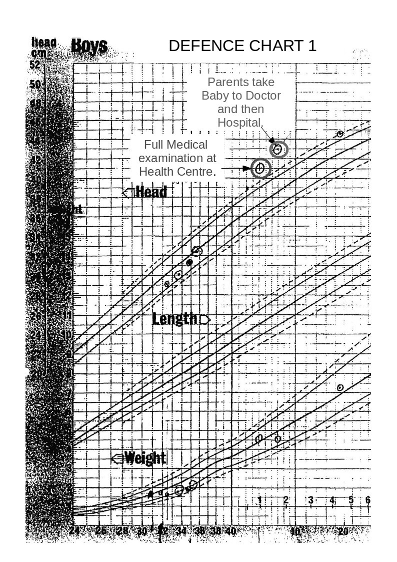
Defence Chart 1
Note; The above graph shows ‘Centile’ curves which are standard guide curves, used to monitor child growth. The centre line on each set of curves is the 50th centile (the ideal curve), the lower dashed line is the 10th centile and the solid line above this is the 25th centile. The upper dashed line is the 90th centile and the solid line below this is the 75th centile.
On the weight curves the measurements show that the child’s weight is actually falling relative to the 50th centile, and is in the danger area. (Whoever entered these point measurements obviously got them wrong at first and then corrected them.)
On the head measurement curves the figures indicate that the head diameter is rising.
In fact the head measurements between the last two made whilst the baby was still in hospital shows that the rise in the period was twice that of a normal ‘term’ baby.
Any doctor should have seen that this was a serious cause for concern.
Was this Senior Consultant Paediatrician lying?
Had this Senior Consultant Paediatrician not seen the charts?
I personally believe that the charts had not been seen.
Note on the above graph the two high-lighted measurements. The measurement taken at the heath centre, 3 weeks before the parents took the child to the doctor and hospital, shows that the head measurement was well above the 97th centile danger level at this time.
This timing is important and will be discussed later.
The defence team received a copy of this chart 5 months after the parents had been charged, despite constant requests for medical records.
The graph below shows a centile graph received at the same time as the previous chart, but obviously created at a later date. You will notice that there are differences.
The head circumferences figures are not now shown for the period from birth to leaving hospital. Also, the plotted point on Defence Chart 1 at the Medical Centre that was shown to be well above the danger level has now been moved down to the 90th centile line.

Defence Chart 2
Strange!
Medical Incompetence or Medical Fraud?
The following chart is on produced by someone on the prosecution team and issued to at least some of the medical/surgical experts in the prosecution team. The family received this attached to the back of a copy of a report issued by one of the medical experts.
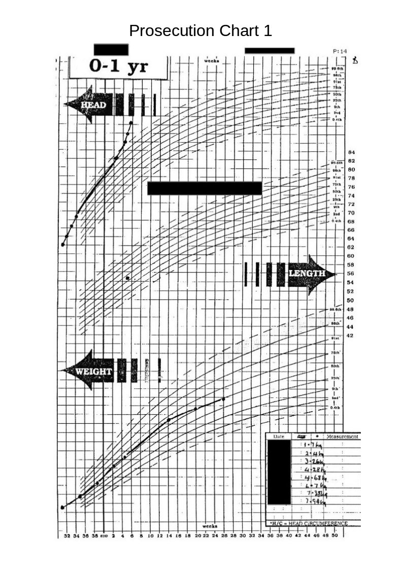
Prosecution Chart 1.
Again we see differences between this and the original charts. The difference between the low weights shown on the original charts and weights shown on this chart, which seem to have mysteriously moved towards the 50th centile, are obviously suspicious.
The head circumference figures (whilst still in hospital), are now lost in a vague area without, any clear definition but indicating that they are on a straight line roughly on the 50th centile.
The following chart, (received with the above chart ) caused me much astonishment and gave me a good, much needed, laugh.
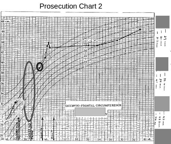 The most obvious thing at first glance was that the chart was a fake made up from two different charts, the long ellipse highlighting the join between the two graphs.
The second most obvious error is that this chart now shows all the head measurements, taken whilst the baby was in hospital, have now magically moved to above the 50th centile. (I would imagine that this is a medical impossibility for a baby being born 9 weeks premature and having a birth weight of 1.7 kilogrammes.)
The third interesting item is that the measurement taken at the health centre and the one taken later at the hospital, have now fallen considerably.
These discrepancies mean whoever produced these charts was grossly incompetent or that these charts produced by the prosecution are deliberate fraud.
Now compare the above chart with the one below.
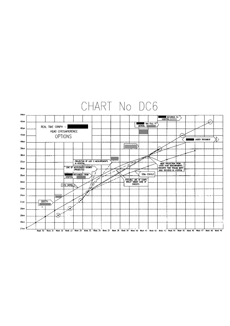
 This is one of the explanatory charts I produced for the defence report.
All of the actual seven head measurements taken in this period are in the correct positions relative to the time scale. The horizontal divisions are in weeks, and the vertical divisions are in 1cm intervals.
Obviously it looks totally different from the previous prosecution one.
Let us look closer at this chart.
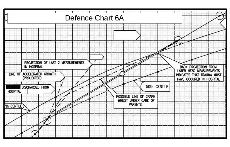 The measurement in the top right hand corner is that taken after the parents took the baby to the doctor and then hospital. The next measurement down is that taken at the Medical Centre two weeks before. All the paediatricians stated that some incident must have happened between these dates (i.e throwing the child about or throwing him onto a hard surface) to account for for the head swelling.
However, projecting a straight line between these these two points backwards, brings it almost exactly to the last measurement made in the hospital.
Author – Brian Williams.